lv bullseye|normal ef bull's eye plot : 2024-10-08 The bull’s eye plot serves as a reconstructional modality based on global and regional longitudinal systolic strain measurements. Its . See more Dieser Automatik-Chronograph ist die 45-mm-Version der turbogeladenen Breitling und beherbergt ein hochleistungsfähiges Manufacture Breitling Caliber 01 in einem ultra-robusten und leichten Gehäuse in Breitlight®: einer .
0 · speckle tracking bull's eye
1 · normal ef bull's eye plot
2 · longitudinal strain bull's eye
3 · bull's eye plot speckle tracking
4 · bull's eye plot
5 · bull's eye mapping
6 · bull's eye echocardiogram
Rolex VS Breitling Full Comparison | Which is the Best? - PrestigeTime.c.
lv bullseye*******The bull’s eye plot offers an intuitive visual overview of the global and regional Lv myocardial function status in various cardiomyopathies with LVH. The bull’s eye longitudinal strain mapping is clinically feasible and the plot patterns derived by a further expansion of this technique in clinical practice provide . See moreThe bull’s eye plot can be acquired either by AFI algorithm or standard two-dimensional (2D) strain algorithm. Both methods are based on 2D STI, with quantitative information generated by measuring longitudinal strain from three apical views (apical . See more
The major technical limitation for longitudinal strain bull’s eye acquisition is the need of high-quality echocardiographic images in standard . See moreThe bull’s eye plot serves as a reconstructional modality based on global and regional longitudinal systolic strain measurements. Its . See more
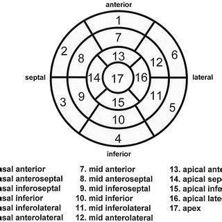
In the left ventricle (LV), the myocardium is arranged in three distinct layers. The sub-endocardium (innermost), the mid-myocardium and the sub-epicardium .The bull’s-eye plot, much like the strain graph, is a visualized version of data collected from strain imaging echocardiography. It gives you information on how well segments of the . “Cherry-on-the-top” sign on STE longitudinal strain bullseye map (preservation of apical longitudinal strain with severely abnormal .
‘Bullseye’ display of regional LV longitudinal strain (with basal regions outermost, mid regions in the middle and apical regions .Aims: Left ventricular (LV) systolic function is a known prognostic factor after ST-segment elevation myocardial infarction (STEMI). We evaluated the prognostic value of LV global.
The longitudinal strain bull's eye plot derived from 2D speckle tracking imaging offers an intuitive visual overview of the global and regional left ventricular . The patient-specific bull's-eye map will help to understand the LV morphology at a glance and to minimize the gap between 3D and 2D LV morphologies because it simultaneously represents patient-specific .normal ef bull's eye plotAt the segmental level, Fig. 3 presents the values for LV ECV, LV circumferential strain, and LV radial strain for the standard 16-segment LV model.
Left ventricular (LV) global longitudinal strain (GLS) is a measure of the active shortening of the LV in the longitudinal direction, which can be assessed with speckle-tracking echocardiography.
The bull’s eye plot in CA patients with normal EF shows a normal or slightly reduced average longitudinal strain, a normal longitudinal strain value at the apex of the LV (bright red), and a significantly reduced strain at all basal segments of the entire LV (pale pink to light red).
In the left ventricle (LV), the myocardium is arranged in three distinct layers. The sub-endocardium (innermost), the mid-myocardium and the sub-epicardium (outermost). The interaction of these layers plays a key role in ensuring stroke volume and hence overall cardiac output.
The bull’s-eye plot, much like the strain graph, is a visualized version of data collected from strain imaging echocardiography. It gives you information on how well segments of the left ventricle wall contract. The contractility is depicted both as colors and global longitudinal strain (GLS) values. Marcello Na. 10th Jan 2022 • 2m read. “Cherry-on-the-top” sign on STE longitudinal strain bullseye map (preservation of apical longitudinal strain with severely abnormal basal and mid-LV longitudinal strain) Characteristic bullseye pattern is likely the most specific sign to rule in the diagnosis of cardiac amyloidosis (but still does not differentiate ATTR vs. AL amyloidosis)
‘Bullseye’ display of regional LV longitudinal strain (with basal regions outermost, mid regions in the middle and apical regions innermost). Left ventricular diastolic function measurements were abnormal.Aims: Left ventricular (LV) systolic function is a known prognostic factor after ST-segment elevation myocardial infarction (STEMI). We evaluated the prognostic value of LV global. The longitudinal strain bull's eye plot derived from 2D speckle tracking imaging offers an intuitive visual overview of the global and regional left ventricular myocardial function in a single diagram. The bull's eye mapping is clinically feasible and the plot patterns could provide clues to the etiology of cardiomyopathies. The patient-specific bull's-eye map will help to understand the LV morphology at a glance and to minimize the gap between 3D and 2D LV morphologies because it simultaneously represents patient-specific segment information on the map with visualization of morphological boundaries.
At the segmental level, Fig. 3 presents the values for LV ECV, LV circumferential strain, and LV radial strain for the standard 16-segment LV model. Left ventricular (LV) global longitudinal strain (GLS) is a measure of the active shortening of the LV in the longitudinal direction, which can be assessed with speckle-tracking echocardiography.
The bull’s eye plot in CA patients with normal EF shows a normal or slightly reduced average longitudinal strain, a normal longitudinal strain value at the apex of the LV (bright red), and a significantly reduced strain at all basal segments of the entire LV (pale pink to light red).
In the left ventricle (LV), the myocardium is arranged in three distinct layers. The sub-endocardium (innermost), the mid-myocardium and the sub-epicardium (outermost). The interaction of these layers plays a key role in ensuring stroke volume and hence overall cardiac output.
lv bullseyeThe bull’s-eye plot, much like the strain graph, is a visualized version of data collected from strain imaging echocardiography. It gives you information on how well segments of the left ventricle wall contract. The contractility is depicted both as colors and global longitudinal strain (GLS) values. Marcello Na. 10th Jan 2022 • 2m read.lv bullseye normal ef bull's eye plot “Cherry-on-the-top” sign on STE longitudinal strain bullseye map (preservation of apical longitudinal strain with severely abnormal basal and mid-LV longitudinal strain) Characteristic bullseye pattern is likely the most specific sign to rule in the diagnosis of cardiac amyloidosis (but still does not differentiate ATTR vs. AL amyloidosis)
‘Bullseye’ display of regional LV longitudinal strain (with basal regions outermost, mid regions in the middle and apical regions innermost). Left ventricular diastolic function measurements were abnormal.Aims: Left ventricular (LV) systolic function is a known prognostic factor after ST-segment elevation myocardial infarction (STEMI). We evaluated the prognostic value of LV global.
The longitudinal strain bull's eye plot derived from 2D speckle tracking imaging offers an intuitive visual overview of the global and regional left ventricular myocardial function in a single diagram. The bull's eye mapping is clinically feasible and the plot patterns could provide clues to the etiology of cardiomyopathies.
The patient-specific bull's-eye map will help to understand the LV morphology at a glance and to minimize the gap between 3D and 2D LV morphologies because it simultaneously represents patient-specific segment information on the map with visualization of morphological boundaries.
At the segmental level, Fig. 3 presents the values for LV ECV, LV circumferential strain, and LV radial strain for the standard 16-segment LV model.
Left ventricular (LV) global longitudinal strain (GLS) is a measure of the active shortening of the LV in the longitudinal direction, which can be assessed with speckle-tracking echocardiography. The bull’s eye plot in CA patients with normal EF shows a normal or slightly reduced average longitudinal strain, a normal longitudinal strain value at the apex of the LV (bright red), and a significantly reduced strain at all basal segments of the entire LV (pale pink to light red). In the left ventricle (LV), the myocardium is arranged in three distinct layers. The sub-endocardium (innermost), the mid-myocardium and the sub-epicardium (outermost). The interaction of these layers plays a key role in ensuring stroke volume and hence overall cardiac output.
The bull’s-eye plot, much like the strain graph, is a visualized version of data collected from strain imaging echocardiography. It gives you information on how well segments of the left ventricle wall contract. The contractility is depicted both as colors and global longitudinal strain (GLS) values. Marcello Na. 10th Jan 2022 • 2m read. “Cherry-on-the-top” sign on STE longitudinal strain bullseye map (preservation of apical longitudinal strain with severely abnormal basal and mid-LV longitudinal strain) Characteristic bullseye pattern is likely the most specific sign to rule in the diagnosis of cardiac amyloidosis (but still does not differentiate ATTR vs. AL amyloidosis) ‘Bullseye’ display of regional LV longitudinal strain (with basal regions outermost, mid regions in the middle and apical regions innermost). Left ventricular diastolic function measurements were abnormal.
Aims: Left ventricular (LV) systolic function is a known prognostic factor after ST-segment elevation myocardial infarction (STEMI). We evaluated the prognostic value of LV global.
The longitudinal strain bull's eye plot derived from 2D speckle tracking imaging offers an intuitive visual overview of the global and regional left ventricular myocardial function in a single diagram. The bull's eye mapping is clinically feasible and the plot patterns could provide clues to the etiology of cardiomyopathies. The longitudinal strain bull's eye plot derived from 2D speckle tracking imaging offers an intuitive visual overview of the global and regional left ventricular myocardial function in a single diagram. The bull's eye mapping is clinically feasible and the plot patterns could provide clues to the etiology of cardiomyopathies. The patient-specific bull's-eye map will help to understand the LV morphology at a glance and to minimize the gap between 3D and 2D LV morphologies because it simultaneously represents patient-specific segment information on the map with visualization of morphological boundaries.
Op Chrono24 vindt u prijzen voor 42 Breitling Ref A13022 horloges, kunt u prijzen vergelijken en voordelig een Ref A13022 horloge kopen.
lv bullseye|normal ef bull's eye plot